|
About this Database
Toxoplasma Full-Length cDNA project
Junya Yamagishi1, Akio Ueno1,
Junichi Watanabe2, Yutaka Suzuki3,
Hiroyuki Wakaguri3, Keiko Toya3,
Sumio Sugano3, Chihiro Sugimoto4,
Makoto Igarashi1, Yoshifumi Nishikawa1,
Xuenan Xuan1,
(1National Research Center for Protozoan Diseases, Obihiro University
of Agriculture and Veterinary Medicine,
2Department of Parasitology, Institute of Medical Science, The University of Tokyo,
3Department of Medical Genome Sciences, Graduate School of Frontier Sciences, The University of Tokyo,
4Zoonosis Research Center, Hokkaido University
)
Summary
|
1. Full-Toxoplasma is a database for transcriptome analysis of Toxoplasma gondii parasites,
which contains our original data produced using the oligo-capping method, i.e., 1)
full-length sequences of full-length cDNA clones and 2) TSS (transcription start site) sequences.
They are shown along with the genome sequences, etc.
|
|
2. Recent introduction of the Solexa sequencers is now revolutionizing quantity and
quality of our data, allowing analyses in depth. Our database contains thus determined
full-length cDNA sequences which require revision of annotated gene structures and
numerous TSS sequences which allow detailed and accurate expression analysis.
|
1.Toxoplasma
1-1. Toxoplasma
Toxoplasma gondii is a protozoan parasite which infects humans
and animals, causing zoonosis. Its definitive host is cats. Many people are
infected by ingestion of oocysts in the feces of cats or eating undercooked
meat containing bradyzoite-stage parasites but few have symptoms.
However, if the first infection occurs during the early pregnancy, the
organisms infect fetus via placenta, causing severe diseases in the brain and
eyes of the baby.
Hydroencephalitis is the most severe form causing mental
retardation, retinitis or retinochoroiditis causing blindness. In addition
immunocompromized people may suffer from its recurrence causing
encephalitis, cardiomyelitis, pneumonia or lymphadenitis.
Toxoplasma gondii is a close relative of malaria parasites and
belongs to Apicomplex kingdom. Comparative biology of these two species is
expected to elucidate naure of parasites.
|
1-2. The Toxoplasma gondii life cycle
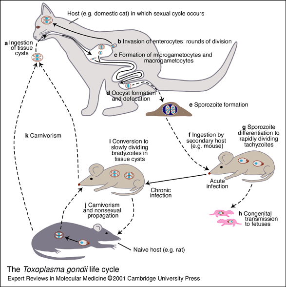 |
T. gondii is an obligate intracellular parasite and its life cycle includes
both sexual and asexual modes of proliferation and transmission. The sexual
cycle takes place exclusively in the intestinal enterocytes of many members of the
cat family (Felidae). (a, b) After ingestion of tissue cysts, the parasites invade the
enterocytes, undergo several rounds of division and (c) differentiate into
microgametocytes and macrogametocytes. (d) The gametocytes fuse to form a
zygote or'oocyst' that is shed into the environment with the cat's faeces. (e) The
oocyst undergoes meiosis, producing an octet of highly infectious 'sporozoites'
that are resistant to environmental damage and may persist for years in a moist
environment. (f) After ingestion (by a secondary host such as a mouse), (g)
sporozoites differentiate into the rapidly dividing 'tachyzoite' form, which
establishes and sustains the acute infection. (h) During the acute infection,
congenital transmission to the developing fetus can occur. (i) In many hosts, a
chronic phase of the disease ensues, as the tachyzoite changes into a slowly
dividing form known as the 'bradyzoite'. Latent bradyzoite tissue cysts persist for
the life of the host, re-emerging occasionally, but do not produce overt disease in
healthy individuals. (j) Carnivorous ingestion of tissue cysts can lead to the
infection of a naive host, allowing for an indefinite nonsexual propagation of T.gondii.
(k) In the cat, this will initiate the sexual cycle. The solid lines indicate
parasite differentiation and the dashed lines indicate modes of transmission
(Ajioka, JW. et al. Expert Rev. Mol. Med. 2001:1-19).
|
1-3. Ultrastructure of a Toxoplasma gondii tachyzoite
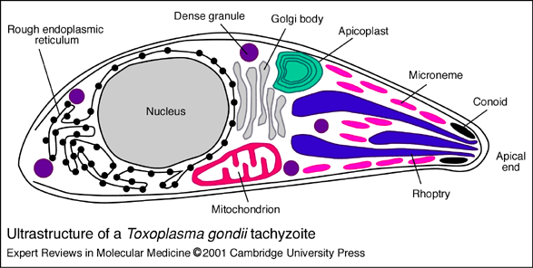 |
The conoid defines the apical end of the parasite and is thought
to be associated with the penetration of the host cell. Micronemes,
rhoptries and dense granules are the three major
secretory organelles, found predominately at the apical end of the parasite.
Microneme proteins are released very early in the invasion process,
facilitating host-cell binding and gliding motility. Rhoptry proteins are
also released during invasion, and can be detected within the lumen and
membrane of the newly generated parasitophorous vacuole (PV).
Dense-granule proteins are released during and after the formation of the PV,
modifying the PV environment for intracellular survival and replication of the
parasite. The apicoplast is a plastid-like four-membrane
organelle containing a 35 kb circular DNA. Most of the proteins functioning
within the organelle are encoded by the nucleus, and are specifically targeted
to the apicoplast. This targeting involves the secretory pathway, including the
rough endoplasmic reticulum (ER) and a Golgi body
situated immediately apical to the nucleus. Targeted proteins have a bipartite
N-terminal extension, consisting of an ER signal sequence followed by a plastid
transit peptide. T.gondii cells have a single nucleus and a single
mitochondrion. It is hypothesised that reliance on the mitochondrion for
cellular metabolism differs according to the life-cycle stage of the parasite
(Ajioka, JW. et al. Expert Rev. Mol. Med. 2001:1-19).
|
- Toxoplasma
- Construction
of Toxoplasma Full-Length cDNA Library
- Current
Status
|